MS-PINPOINT · UCL Queen Square

Turning routine hospital data into answers about multiple sclerosis.

We build the computational tools that read the scans and records hospitals already hold — across many hospitals, internationally — so that what happens to one person with MS can inform the care of the next.

Eight axial slices through the MNI152 brain template, ascending from the cerebellum to the vertex
Sagittal spine MRI with Spynth spinal cord segmentation shown in colour by vertebral level

Two studies

One running today. One starting in 2027.

Running now

MS-PINPOINT

Sites
12 hospitals
Based at
University College London

Bringing together routine imaging and clinical data from twelve hospitals to work out what actually predicts how multiple sclerosis progresses in an individual.

This is the study with an opt-out: if you are a patient at one of the twelve hospitals, it is the one to act on.
Starts July 2027

DREAMS

Sites
16 hospitals
Based at
King's College London

Our next study, newly funded and beginning when the group moves to KCL. It widens the network to sixteen hospitals and takes a new direction: AI that understands free text and conversations with patients, for a future in which people hold a central role in their own care, supported by doctors both human and AI.

It has not started, so there is nothing to opt out of yet and no data is being collected for it.

If you are a patient at one of the twelve MS-PINPOINT hospitals and do not want your data included, the opt-out above is the one you want. DREAMS has not started and has no opt-out yet.

Software

MindGlide reads the scans hospitals already have.

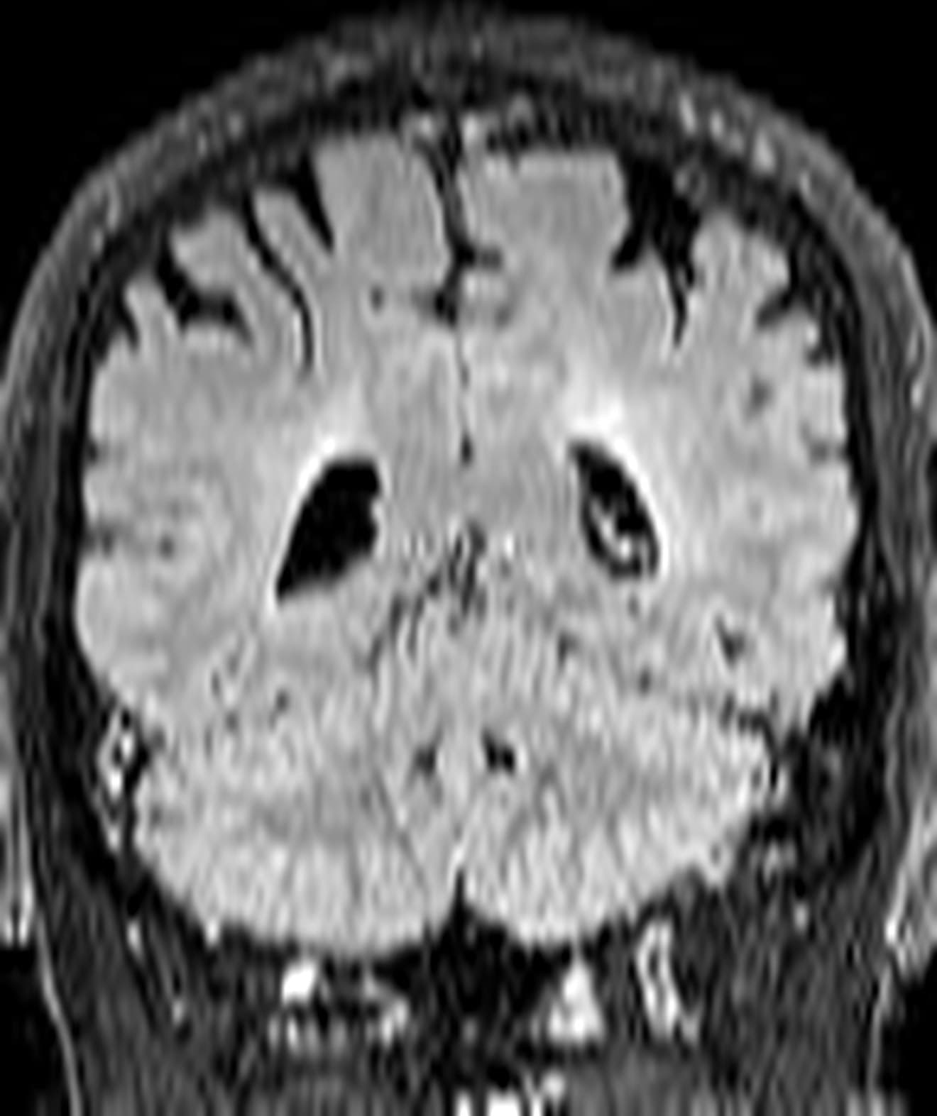
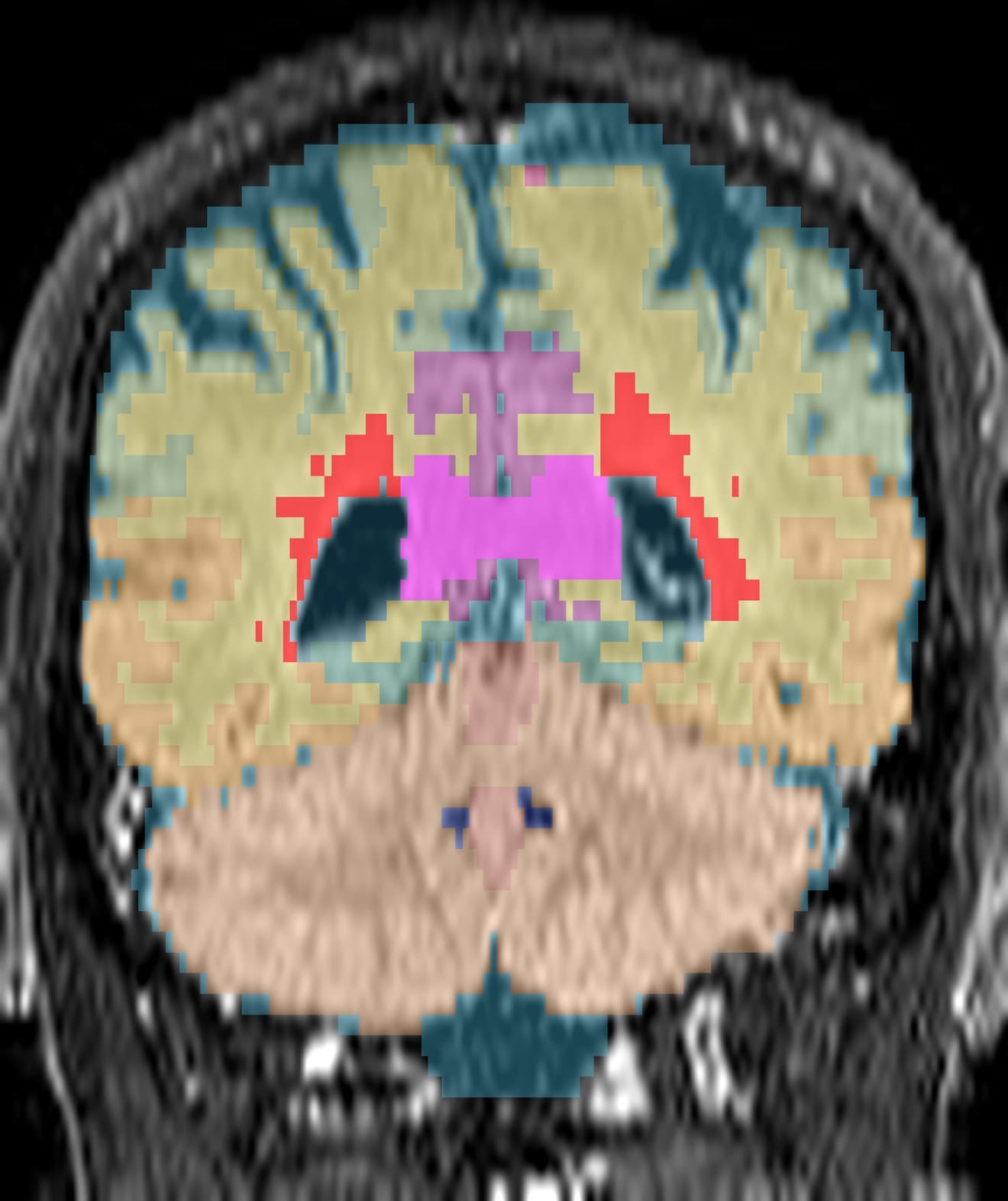
Hospital MRI archives hold decades of imaging that was never analysed at scale, because the scans were acquired for clinical care rather than research. MindGlide is our deep-learning tool for measuring them anyway — turning an archive into a dataset.

A coronal FLAIR MRI slice of a brain affected by multiple sclerosis, as the hospital acquired it: bright patches sit around the ventricles, but nothing has been outlined or measured.
The same slice after MindGlide has read it: each brain region is shaded a different colour — grey matter, white matter, cerebellum, corpus callosum and the ventricles — and the multiple sclerosis lesions are picked out in red beside the lateral ventricles.
Raw FLAIRMindGlide
Colour key for the segmentation: cerebrospinal fluid, the third, fourth and fifth ventricles, the lateral ventricles, cerebellum and cerebellar vermis, temporal lobe, corpus callosum, white matter, frontal, limbic, parietal and occipital grey matter, and lesions.
Drag the handle — or focus it and use the arrow keys — to wipe between a routine hospital scan and MindGlide's reading of it. This is an illustrative research rendering, not a clinical diagnosis.